|
Vision Care 3 years to 10th grade
Slide 1.

Vision care of school children has changed
during the last fifteen years when children with different disabilities
have become integrated in local schools. Instead of basic vision
screening that has been the content of vision care given by the school
nurses, we now participate also in the transdisciplinary assessment of
vision for IEPs of children with special needs.
This lecture was given in Las Vegas, April 28.
- 29. 2004, as a part of discussion on development of vision services
for school children. A discussion like this should be repeated after a
few years so that all nurses are aware of the principles of screening,
of individual assessment of vision and of referral practises. Deeper
understanding of the content of the services and of the role of vision
in the development and functioning of children leads to effective use
of examination techniques and better quality of services. In several
instances this improved quality of working saves time and money and
therefore it interests also the administration of the services and
insurance companies.
Although variations in vision screening are great, many central features are common in all countries. The discussions of these two lectures and in the short overview of vision services for infants and toddlers (0-3 years) get their special flavor from the traditions in Nevada and in Finland. In both places vision screening has defined its goal as follow-up of development of vision and early detection of deviations from normal development, much the same way as length, weight and hearing are measured exactly. Thus the vision services cover more than amblyopia screening. In Nevada there are also long traditions of participation of school nurses in assessment of vision of children with special needs. Because of these goals, measurement of visual acuity is done until threshold and nurses have experience in assessment of visually impaired children.
Since the discussions during these lectures were found interesting, they are made available to a larger group of workers in the field to further activate discussion and comparison of different vision services.
INTRODUCTION
Slide 2.
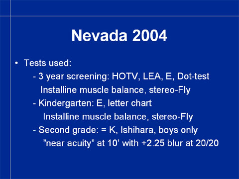
In Nevada, the techniques used in vision screening between 3
years and the second grade are now:
In the screening of the three year old children:
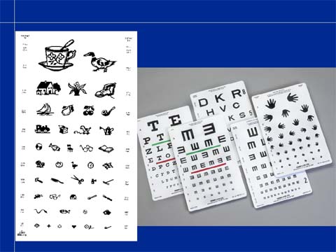
- visual acuity tests: HOTV, LEA, tumbling E and Dot-test
- Insta-Line muscle balance test
- Stereo-Fly, Stereo-Reindeer
In the screening of children in kindergarten:
- visual acuity tests: E and letter chart, Sloan letters
- Insta-Line muscle balance test
- Stereo-Fly, Stereo-Reindeer
In the screening at the second grade: the same tests as in
kindergarten and color vision screening with Ishihara test, only boys.
Slide 3.
The use of several visual acuity tests often means that a
child is tested with three different tests in the three test
situations. Since each test measures vision slightly differently, the
results are not comparable. Normally there is an increase in the visual
acuity values by 1-2 lines between these measurements when the same
test is used in each test situation.
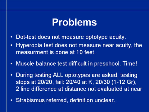
The Dot-test (add manufacturer) is not a visual acuity test
but an eye-hand-coordination test. The child is asked to point to the
dot. Thus the visual task is to localize where there is a dot on the
white surface, a parietal lobe function, whereas visual acuity measured
with optotypes is a recognition function of the inferotemporal lobe.
(For structure of visual pathways see Section 4).
Hyperopia test was introduced when the services were
criticised for lack of near vision testing. Hyperopia test does not
assess quality of near vision but may reveal higher than usual
hyperopia. If hyperopia does not cause decrease in visual acuity and/or
esophoria it does not need to be corrected and therefore hyperopia test
is not particularly useful. This has also been the experience of the
nurses. Since the test situation is experienced difficult and time
consuming, its use could be discontinued. The same applies to the
muscle balance test.
During the measurement of visual acuity, all optotypes on each
line are asked. This requires more time than if only the first/last
optotype on each line would be asked. The pass/fail criteria could be
re-evaluated.
Only the picture of the Stereo-Fly or the Stereo-Reindeer is used in screening. Since there are in these tests also stereo images that require better stereo resolution and thus depict the level of binocularity better, and since many children can respond to these pictures, they should be used in the examination. Stereo tests are expensive and therefore they have not been accepted in many countries as a part of amblyopia screening. Since all nurses in Nevada have a stereo test, either Titmus Stereo-Fly or The Bernell Stereo-Reindeer, their use is recommended.
The guidelines of referral in case a child has strabismus
should be defined. Since strabismus may lead to development of
amblyopia, it needs to be included in the list of techniques to detect
amblyogenic factors.
Slide 4.
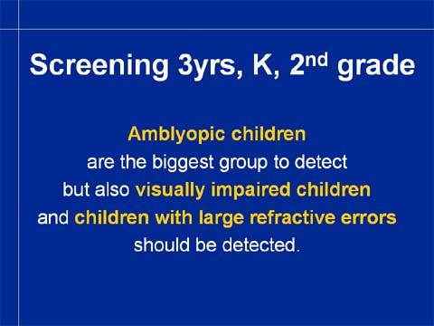
In the care of children of preschool age and
still on the second grade level, amblyopia is the most important
deviation from normal development to be detected. Visual acuity tests
that are used in screening of amblyopia detect also a small
number of children with decrease of visual acuity in both eyes, i.e. visually
impaired children or children with large refractive errors.
Among normally developing children visual impairment and large
refractive errors are rare. Most cases of visual impairment are
congenital and 60-70% of visually impaired infants and children have
other impairments and disorders, so they should have been diagnosed
before the screening at the age of three years.
Vision screening, like all screenings,
concerns only symptom free healthy children.
If a child has intellectual disability,
behavioral problems, motor problems, hearing impairment or any syndrome
or disorder, the child is assessed with tests that are specific to that
particular group of children. Most of these conditions are diagnosed
during the first year of life and thus also assessment of vision by a
paediatric ophthalmologist and by an early intervention team are
performed as a part of the initial investigations. There should be
information available from these earlier clinical investigations of
each impaired child.
Healthy children should have information on
their early development: check on red reflex and structure of the eyes
at birth, eye contact at the age of 6-8 weeks, normal milestones in
interaction, reaching for and grasping, recognition of family members
and peers and no or minor alignment problems of the eyes. If they are
not recorded earlier, it is wise to ask for this information when the
child is seen for the first time at the age of three years or later.
Slide 5.
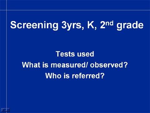
In discussion of vision screening, we cover tests used at different ages, witch functions are measured and observed, and who is referred to further assessment.
VISUAL ACUITY TESTS
Slide 6.
In the mid-70s, paediatric visual acuity tests were poorly calibrated and contain optotypes that require many other functions but recognition of the optotypes. It was therefore I started to develop visual acuity tests that would function better in both screening and assessment in 1976. The designing of the tests followed the advice given by the Department of Psychology at the Helsinki University: in order to have a sharp threshold of the measurement, the tests should be based on optotypes that blur equally.
Slide 7.
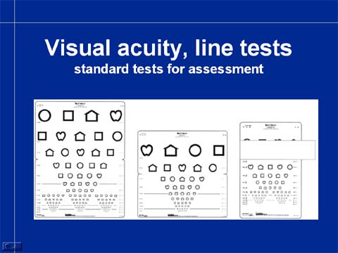
The four optotypes that I designed were the
first four paediatric optotypes that blur equally. This is apparent if
you look at the tests at a distance where the lowest lines are blurred.
You see the optotypes clearly until a certain line; below it all
symbols are blurred to circles. The threshold is very sharp (not as in
some letter and number charts where certain letters or numbers are
clearly seen two lines below the line where other optotypes are
blurred).
It was thought in 1970s that a distance visual
acuity chart for 3-4 year old children, another for 5 year old and
older, a near vision test and single symbols tests for distance and
near would be all that would be needed for good vision services. Later
I have designed several other tests to meet the needs of different
clinical test situations.
Slide 8.
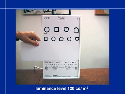
Translucent tests with back illumination have
become standard in many states and countries because the luminance
level is always the same and the lightboxes are easy to move from one
screening location to the next. In Nevada the Insta-Line is the
standard test and thus development of screening is based on it. Its
test layout will be redesigned to allow use of five symbols on each
line and the size of the symbols will be until 20/10 so that there will
be at least one line that the child does not see. This allows measurement
until threshold and thus makes it possible to detect a difference of two lines between the eyes.
Slide 9.
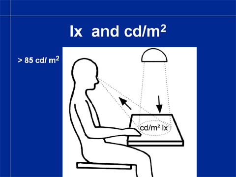
Visual acuity values become higher when
luminance level increases until approximately 85 cd/m2. At higher
luminance levels the visual acuity values first remain the same as at
85 cd/m2 and then at very high luminance levels dazzling decreases
visual acuity.
Slide 10.
The near vision test is a miniature copy of
the distance visual acuity chart.
Slide 11.
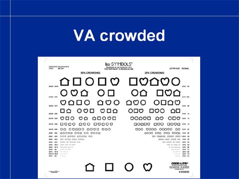
On the reversal side of the near vision card
there are tests with less space between the optotypes. These tests
measure crowding phenomenon more sensitively than the classical
near vision test on the front of the test. Increased crowding is
typical to amblyopia and several types of impaired vision and means
that a child cannot keep optotypes and other details apart but they
glide partially on top of each other. “They hug each other”, as a
little patient of mine once described the phenomenon.
Slide 12.
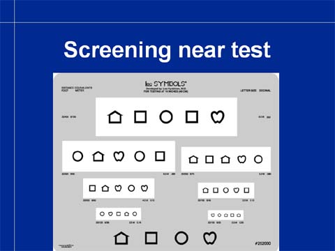
The standard near vision test is somewhat slow
in screening. Therefore there is this screening version of the line
test with more space between the lines. The test is easier for the
child and the tester because it is easier to know, which line needs to
be read.
Slide 13.
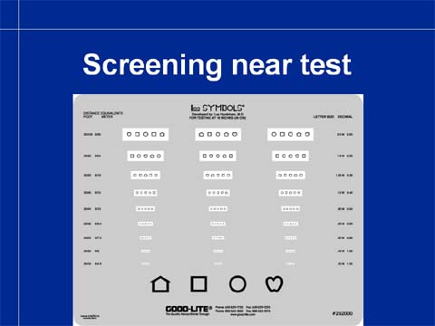
The smaller optotypes are in three well-separated lines so measurement until the threshold level is quick. The grey background dazzles less than the white background in assessment of childer with visual impairment.
Slide 14.
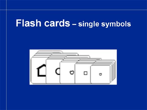
Children who cannot perform in the line test
situation are tested using single symbol tests where there is one
single symbol on a large white surface. Such single optotype is much
easier to recognize even if fixation and accommodation are not normal.
Therefore visual acuity values of amblyopic eyes measured with single
symbols can be 2-3 lines better than those measured with line tests of
the same optotypes. Even one line difference in single symbol tests
might mean beginning amblyopia.
Slide 15.
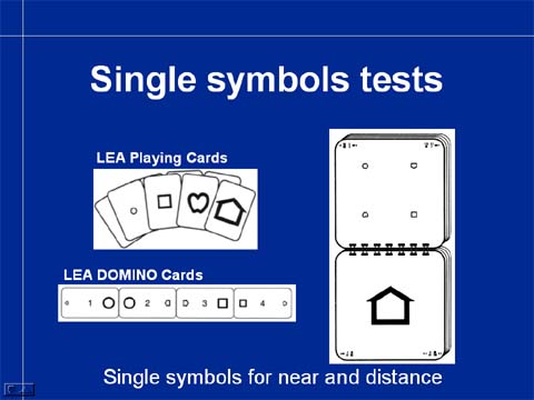
Single symbol tests are also in form of a
small book for distance and as playing cards for near. The smallest
symbols are in the LEA Domino Game that was designed for training of
amblyopic eyes.
Slide 16.
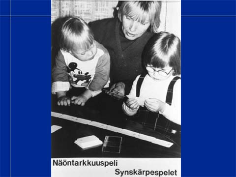
In this picture you see the three year-old
girl playing domino game with the LEA Domino cards. There is patch on
the right lens of the glasses to make the child to use the amblyopic
left eye. A younger sibling observes the play situation and learns the
symbols quite early. This is important because he might be in danger of
developing an amblyopic eye and therefore follow-up of the development
of visual functions should include measurement of visual acuity as
early as possible.
Slide 17.
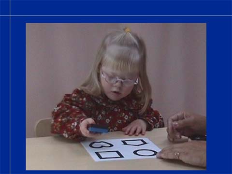
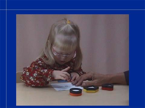
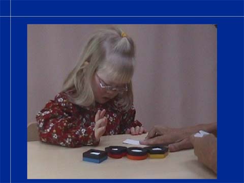
For training of the test situation I have
designed the LEA Puzzle that is useful in training young children and
children with special needs. We all learned match colors before we
could match forms. Therefore there is the colorful side of the puzzle
where the child can place the puzzle pieces by matching the colors. At
the same time the child is playing with the forms (in color) and learn
the locations of the different colors and forms on the puzzle board.
With this training children can be tested with single symbols test at
near before they are 18 months old. The youngest have been 13 month
old. If also covering of one eye at a time is trained, as a part of the
play, screening of visual acuity is easy and quick.
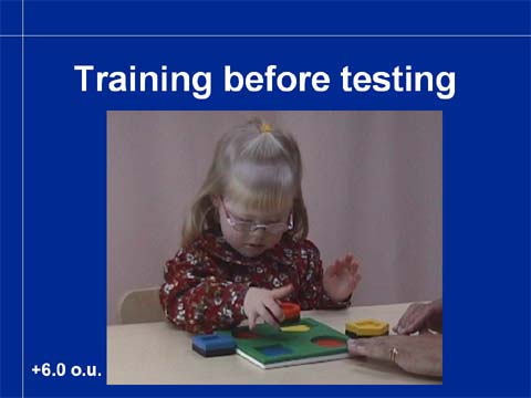
Slide 18.
Like this little girl with Down Syndrome, many
children with special needs require a shorter or longer time to train
for measurement of visual acuity with optotypes. Some children need to
train with the puzzle board at the first grade. Since this activity is
typical to toddlers and young children it can be included in their play
situations in the day care and at home.
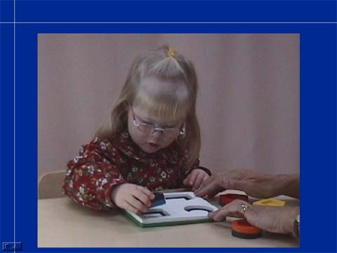
When the child masters the colorful side
of
the puzzle, the black-and-white side is introduced. When the child can
play with the black-and-white forms, a much more difficult step in
development is trained: comparing a concrete object with its picture,
first the same size, then smaller and smaller. At this stage we are
already measuring visual acuity with single symbols.
Slide 23.
If the development of the concept of
pictures
representing objects is difficult, drawing around the child’s hand,
foot, favorite toys, small kitchen utensils etc. is helpful. After that
drawing around the puzzle pieces may make the comprehension of these
slightly more abstract pictures possible.
Slide 24.
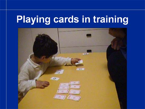
Playing cards can be used as a part of occupational
therapy
and function as a fun test of single symbol acuity. By measuring the
distance that the child uses to look at the symbols (preferably with
your hand instead of a ruler) and knowing the M-size of the smallest
symbols correctly recognized, the visual acuity can be calculated by
dividing the distance in meters by the M-size. For more information,
see the Instructions Section.
Slide 25.
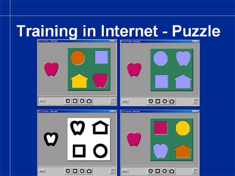
Children can play with the symbols also on
the
Internet on my homepage in Section "Games". This is a
neuropsychological test where one can observe the child’s ability to
match colors, forms in color and black-and-white forms.
Slide 26.
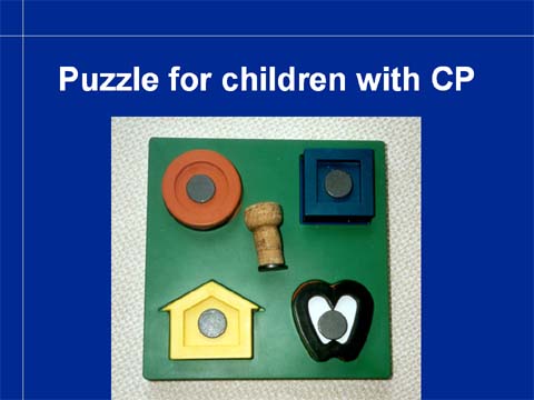
When we test children with CP, the LEA 3-D
Puzzle
needs to be modified by using small magnets on the puzzle pieces and on
a champagne bottle cork to allow the child to move the puzzle pieces.
Slide 27.
When the LEA symbols tests are used, the
same
tests can be used in vision screening of healthy children during
screening at 3 years, kindergarten age and at the second grade. Then it
is possible to notice changes in visual acuity values. Normally, visual
acuity increases by 1-2 lines between each measurement.
If the families and day care receive
information materials on the importance of early vision screening and
the children train the test situation, measurement of visual acuity
becomes a quick and accurate procedure.
There is variation in the functioning
of
children in their performance during vision screening. Although a
great majority of children has exactly the same results in repeated
measurements, still at the age of seven years some children show a
variation of plus/minus two whole lines in their visual acuity when the
measurements have been made in the same room and with the same
instruments. Therefore visual acuity values should e measured twice
before making the decision to refer. (I have learned that it is the
rule also in Nevada.)
OCULOMOTOR FUNCTIONS & STRABISMUS
Slide 28.
In the vision screening before the school
age
and at the second grade we look for signs that could mean deviations
from normal in the vision development.
Fixation and following/tracking are
quick test situations that may reveal difference in the use of the two
eyes. We look for unsteady fixation or fixation nystagmus during
monocular fixation, irregular following movements instead of smooth
pursuit movements.
Strabismus often causes amblyopia
in
the strabismic eye, especially in inward squint. Therefore testing for
alignment with cover test is a valuable test.
We measure visual acuities with line
tests
because it is an effective way to detect amblyopia. Normal visual
acuity does not exclude cerebral visual impairment, brain damage
related impairment. Therefore a list of the most common symptoms of CVI
could be used as a part of vision screening to diagnose also CVI
earlier than it is diagnosed presently.
Slide 29.
For the testing of fixation we need an accommodative
target.
I have designed my fixation stick based on the studies of Robert Fanz,
who showed that picture of face with a diameter of 5cm/2 inches
interests a normally sighted baby at the age of three months and later
so that fixation and following movements can be observed. The two
smaller pictures, giraffe and bunny, are used in the examination of
children at the age of 3 years and older because they are interested in
observing small details of the pictures.
If the fixation stick does not interest a child, we use
different toys to catch the child’s attention for long enough a moment
so that cover test can be performed. My best toys are a baby food jar
with a plastic figure glued on the lid and with “snow” flakes in water.
Another good toy is a toy mobile phone in the assessment of infants.
When the child is playing with any of these toys, he usually does not
notice cover in front of an eye if both eyes are normally sighted.
Slide 32.
In screening we usually use only direct
cover
test because alternating cover has been experienced confusing. With the
direct cover test we can observe both tropias and phorias.
Slide 33.
A few examples of cover test findings
might
make the test situation easier to understand. This first variant of
inward squint, esotropia is the most benign type. The child can fixate
with both eyes equally well, switches from one eye to the other eye
spontaneously, so there is no greater risk of development of amblyopia.
However, also this child needs to be referred so that the refractive
status and central vision of both eyes are assessed more carefully. At
the end of the sequence the infant’s gaze happens to fall on his father’s face and therefore remains in its inward position. His father
namely responded to the eye contact by saying something and was more
interesting a target than my fixation target.
Slide 34.
This child has constant esotropia of the
right
eye. The eye is turned inward but fixates centrally when the leading
left eye is covered. Fixation is stable. This eye may be amblyopic but
the child uses the eye still quite well.
Slide 35.
You need to concentrate on watching
carefully
when the right eye is covered for the first time. Fixation is unsteady,
searching. Also during the repeated cover fixation is unsteady so it is
likely that this eye is amblyopic, which it was found to be. However,
it has recovered well during therapy.
Slide 36.
In this case the camera angle has been
chosen
to be oblique so that the movement of the eye behind the cover can be
seen well. During assessment the movement is quite well noticeable
behind the cover but the camera loses much of the clarity of the image.
Behind the cover the leading, dominant eye turns in and immediately
takes fixation when the cover is removed.
Slide 37.
If exotropia, outward squint lasts only a short period, a few minutes when the child is tired or is sunken in his thoughts, it does affect the development of binocularity in children who have their eyes aligned most of the time and have already developed strong binocularity with good stereovision. (Intermittent exotropia is a common finding in the listeners toward the end of lectures.)
Slide 38.
At this address in the Internet you find
the
following animations for training of the cover test.
Slides 39 - 43 are in the PowerPoint lecture only.
Slide 48.
It is worth watching these slides over and over again. In order to make the training effective, the slides are here without the headings and in the PowerPoint presentation you have both variants. (The animations will be on the PowerPoint and on this text on the CD.)
Slide 49.
Hirschberg’s test or corneal light reflex
test
is a much-used quick test to detect exo- and esotropias. It does not
reveal small angle tropias. If reflexes are symmetric and central the
eyes are aligned or the angle of strabismus is small. If the corneal
reflex is nasally, the child has exotropia. If the reflex is
temporally, the child has esotropia.
VISUAL ACUITY BASED SCREENING
Slide 50.
Visual acuity based screening has been the backbone of
vision
screening in the State of Nevada and many other states and countries
for more than thirty years. It has nearly uniform structure in most
programs. It can be started at the age of three years but at that age
too many children react negatively to the covering of an eye, whereas
at four years of age, nearly all children accept covering of the eyes
in turn. The success of testing can be greatly improved if the
three-year-old children have played covering-the-eye-games and played
with the 3-D LEA Puzzle and the Playing cards. During these games the
teachers and parents may have noticed that a child allows covering of
one eye but not the other. This should be known as one of the signs of
too big a difference in the function of the two eyes and thus a
diadnostic sign for referral.
WHAT EXACTLY ARE WE TRYING TO FIND WITH THE VISUAL ACUITY SCREENING?
Since we have already detected children who have tropias
either in the Hirschberg’s test or with the cover test, the children to
be found with the measuremnt of visual acuity are
- Children who have so small angle strabismus
that it
has not been noticed in the previous two tests, but which has lead to
asymmetric development of visual acuity or
- Children whose eyes are aligned but have unequal
refractive errors and therefore also unequal visual acuity; Anisometropia may have caused amblyopia in the eye with more blurred image but this is not very common at the age of four years. If the child has symmetric corrected visual acuity and normal stereo acuity, the family may decide not to buy glasses but check visual acuity at the kindergarten after a few months hoping for emmetropisation of the eye with higher refractive power.
- Children with decreased visual acuity in both eyes
either
because of large refractive errors or visual impairment of
both eyes due to disorders in the eyes, in the visual pathways or
in the higher visual functions. This is the smallest group because
nearly all visually impaired infants and children are nowadays
diagnosed early among the children with other impairments (60-70% of
visually impaired children have at least one other impairment or
disorder or chronic illness). We need to be aware of the group of
children with hereditary retinal disorders who may have been symptom
free until 3-5 years of age. Most of them have symptoms of beginning
difficulties in dim light so this should make the persons taking care
of the child to start questioning. Also, there is the small group of
children with storage diseases who may have had perfectly normal
development until 2. – 5. year of life and then lose some of their
visual acuity and often at the same time develop behavioral problems.
Therefore children who “do not concentrate” or “do not co-operate”
should have their sensory functions carefully examined before they
start receiving any therapies. This, not because the therapies wouldn’t
be helpful, BUT because the child’s condition needs to be diagnosed.
It is generally accepted that the difference between the
visual acuity values at distance needs to be at least two lines to
warrant referral. The referrals can be further restricted to cases
where there is the same difference also in the visual acuity values
at near. Since myopia is rather rare in the preschool population,
this addition to the measurement battery does not notably affect the
screening before school age, whereas it markedly reduces the number
of wrong referrals in the teen-age population. Therefore it should
interest the administration of the services.
(When I started as the ophthalmologist in charge of the schools in Helsinki in 1983, the pass/fail limit was visual acuity 20/30 in one or both eyes. Therefore more than half of referred children had so mild myopia that it did not require correction. Several parents were angry asking why they had been asked to take half a day off from work (unpaid) to come with the child to my office when nothing needed to be done. I felt that it was an important question and introduced the use of near test to find out that a child who earlier had had visual acuity 20/16 in both eyes and now had 20/16 and 20/30 at distance and 20/20 in both eyes at near, that (s)he did not have anything else but beginning myopia. Namely, if the child has 20/20 visual acuity at near in both eyes, then only myopia can be the cause of decreased uncorrected distance visual acuity. This decreased the number of referrals in Helsinki to one half of what it had been and we got time to assess all special needs children at their schools together with the school nurse and the assistants whom the children wanted to be with them during the assessment.)
It is important that health care services do not refer
children who do not need to get glasses because there is a risk that a
child may get very weak glasses that remain mostly in a drawer. This
happens in all countries, especially if the doctor has any economic
interest in selling the glasses.
In the control of the quality of referral services, it is
important to get reports from the doctors. Also, if children are asked
to have follow-up by the eye doctor for no good reason (it has happened
that nearly all children were asked for follow-up in six months) the
situation needs to be discussed with the local eye doctors and the
administration.
VA SCREENING TECHNIQUE
Slide 51.
The child needs to be able to match or
name
the symbols. Matching requires the concepts similar/different, which
can be trained. When naming is used ANY NAME is OK. Children must not
be corrected, even if the name might be as strange as “bunny ears” for
apple. The name may also change. The house is in the larger sizes house
and then becomes a “dog house”; similarly apple may become berry in the
smaller sizes.
Slide 52.
The order of measurement that I have found
to
function well, is following:
- Binocular near visual acuity, the most important VA for
learning
- Binocular distance visual acuity, the functionally
important VA value at distance
- Both monocular visual acuity values at distance,
usually
the RE first, then LE, except when the child reacts clearly negatively
to covering of the left eye (which may be the non-amblyopic eye).
- If there is difference between the monocular values at
distance then also monocular values at near are measured.
Slide 53.
Children perform best in vision screening
if
it is fast and easy. Therefore it is important to make it
child-friendly:
Point on the symbols on the uppermost line
and
decide with the child, which names the child wants to use. Respond with
a low key “Those are nice names”.
Ask the child what is the first symbol on
each
or each other line. When approaching the threshold ask also “And what
is next to … (the name that the child used for the first symbol). This prepares the child to read along the line. Do NOT point, it makes fixation easier. Rather cover the line above the line to be read.
When the child makes an error or
hesitates,
move up one line and ask the child to read the whole line. Support a
young child by saying “And next to the…” after each symbol. If the
child has answered incorrectly, say “And after that there is?” This helps the child to concentrate on looking on the line and thus keep the eye correctly focused. If the child looks at you, focusing is likely to change, so if the child does see symbols on the line where the symbols were seen, go back one line and ask the child to name the first symbol and then go back to the line below. Do not let your voice get tense,
Visual acuity is the value of the line, on
which at least three out of five symbols are read correctly.
When writing down the result, the most
exact
way is to write the number of symbols that were read correctly; i.e. if
20/25 line was read correctly and one symbol on the 20/20 line, the
result is 20/25(+1).
In Finland, we use the same rule as is common in Nevada: if a child does not get a normal result, then the measurement is repeated either by the same person or by another person, whom the child knows well, usually the school nurse of his/her own school. It is important to know that visual acuity values of normal well functioning children may vary two lines worse or better than the value measured in the same room with the same instruments a week or two before. The number of these children is not great but they are there in all populations.
Slide 54.
Covering the eye is often difficult.
Therefore
we have tried to develop different ways of covering. One of them is
this special frame with straight sidebars, the other much-used
technique is having two pairs of sunglasses with one lens removed in
each and the other covered.
Slide 55.
In the INSTA-LINE visual acuity can now be measured until 20/20. It will be changed so that the smallest symbols/numbers/letters correspond to 20/10 and there are 5 symbols on each line. The muscle balance test and the hyperopia test that have been critizised here will not be integral parts of the test system, but will be available.
Slide 56.
Vision screening should detect all different types of
visual
impairments. The rapidly increasing number of young children with
cerebral visual impairment, CVI, needs to be diagnosed earlier than
today. Too many of these children come to school without early
intervention services and enter school system that is not prepared to
take care of them.
Present visual acuity screening detects only a part of them, so we should add to our screening techniques a questionnaire on the most common symptoms of CVI. Also we should follow the recommendation that all infants with other impairments and disorders need to be assessed, not screened, preferably as a part of the initial investigations also for their sensory functions; most often during the first year of their life. This would be the most effective way of getting all visually impaired infants within the early intervention services. It requires further education of both paediatrician, paediatric neurologists, ophthalmologists and audiologists.
Slide 57.
At this moment we wanted to have a break.
Slide 58.
This is the question for the second part
of
this discussion.
Slide 59.
This is the present situation in Nevada.
There
are three mandated screening times during the period when the majority
of children who are going to become myopic start to move on the myopic
side.
Slide 60.
Screening in school age after the second
grade
is totally different from vision screening in preschool and still on
the second grade. We are no more searching amblyopic children or any
other treatable condition but children who have not themselves noticed
that they could benefit from using glasses. As it is written in the
slide: the question is not a disorder or disease but VISION COMFORT at
school.
Since the question is of vision comfort,
we
need to define what is needed for comfortable seeing at school.
Children with special needs have such a great variation in their vision problems that no screening functions, Similarly as before the school age, these children need to be examined by their ophthalmologist/neurologist/audiologist and followed by their team at intervals that are decided individually for each child. Children with CVI are often the least understood students at all schools because people cannot understand and believe that a child may function normally in most visual tasks and yet be blind in other visual tasks.
Slide 61.
The technique used in measurement of
visual
acuity is the same as in preschool screening. Since the children should
not be referred too early or too late, we need to define what is needed
for good functioning at school. For adult person 20/40 visual acuity is
accepted as good enough for driving, therefore one might think that it
could be an acceptable pass/fail line at schools as well. However, if
the student is allowed to sit in front, a visual acuity of 20/60 will
be enough. On the other hand, if a student is sitting in the last row
of a class of 40 students and the handwriting of the teacher is not
good, even 20/25 will be too low. This means that we need to know the
schools and use common sense more often than we are used to.
Now the near vision test becomes important. If there is a drop in the visual acuity value (compared with the previous screening) also in the second measurement, we measure the near vision acuity. If the near vision values are normal and close to the previous ones or better, the change in the distance vision is related to myopia. The child has good vision, is only a bit myopic. Myopia is not a disease that needs to be treated but may cause discomfort, so we need to know whether the child has discomfort in reading from the blackboard or whiteboard. If not, (s)he will not buy glasses or use them if they are bought but rather squeeze to see the smallest details. Squeezing is not dangerous and at their age they do not get wrinkles. Their myopia will not develop faster or more slowly if they don’t use glasses.
Summa summarum, there isn’t one clear
pass/fail line but the comfort line.
Somebody asked: what if one eye is more
myopic
than the other? Say that the one eye has visual acuity of 20/25
(previous 20/12) and the other eye 20/100? This is no problem at all.
Usually the more myopic eye is preferred in reading and the less myopic
eye when looking at distance. Thus the student needs to accommodate
very little when looking at near, sees well both at near and at
distance – and will do so for years, in some cases through their whole
life and will never need glasses. Again, comfort is the deciding line.
As long as the child has no discomfort (s)he is not referred. If
(s)he has discomfort (of any kind), either it is treated by the school
doctor or the child is referred independent from what the visual acuity
values are.
Somebody said that your rules ask you to
refer
all children who have VA values below a certain limit. This is
something what I would like to see you reconsidering. If health care
personnel refers students who do not need glasses to doctors who a
likely to give them weak glasses that the student does not need, then
the personnel does not function as proper health care personnel.
Slide 62.
If it is impossible to define a VA value
for
pass/fail in case of myopia, it is even more difficult to define a VA
line for referral in case of hyperopia or astigmatism. If the student
is symptom free and has no esophoria, I would not like to see
him/her in my office.
Question: But if the student has
headaches? –
A student with headaches shall not be screened but treated. The causes
of headache are many; lack of glasses is not the most common but the
breakfast. We have as a rule that a student must eat, sleep and be
active outside according to the recommendations given by the school
nurse for two weeks, and the possibility of poor ventilation of the
sinuses, disturbances in the bite and the neck-shoulder problems have
been examined and ruled out, before a referral to the consulting eye
doctor can be considered. Now a day it is common that parents would
rather have their child treated by a doctor than accept the trouble of
bringing up the child to follow healthy habits.
Slide 63.
The tests used for screening are based on
the
LEA Numbers or the EDTRS letters. Children older than seven years like
numbers best. Letters add one more brain function, reading to the test
situation. Numbers are also internationally acceptable, whereas the
Roman letters are used in a limited area. (In Finland we are aware of
this fact because east of our eastern border Cyrillic letters are used,
farther east hundreds of different languages with their different
characters are the reality. The LEA Numbers were designed for
international studies in occupational health.)
I would like to stress once more that the
use
of the near vision test helps us to find out that there is no other
change but beginning myopia when there is a drop in the distance visual
acuity value.
Someone brought up screening of children
who
have glasses. Of course we measure their vision but their care is not
based on the screening pass/fail but when they feel it worthwhile to
pay for another pair of glasses. To clarify this point I would like to
tell a true story of a Finnish bright boy who got his second pair of
minus lenses. He said to his mother:” Without my glasses I see that
there is a tree on the yard. With my old glasses I see all the
branches, even the thin branches. With the new glasses I see every
single leaf. Do I need to see every single leaf?”
This brings us to the next slide and to
the
question “What is important in the visual information?”
Slide 64.
This slide depicts three photographs. The
picture on the left is the original photograph. The picture in the
middle was kindly made for my teaching by Lamberto Maffei in 1981. He
filtered, removed the fine lines, so in this picture there are no lines
that correspond to high visual acuity. The picture to the right
contains only the fine lines. We all experience that there is more
useful visual information in the picture in the middle than in the
picture to the left. This is one of the most important slides of this
lecture. If you remember that important information for visual
functioning is NOT close to 20/20 but 20/200 and at low contrast, then
you take more calmly the small changes in the high contrast visual
acuity values that you measure.
Slide 65.
Visual information at low contrast levels
requires broad lines, large forms to be transferred through the visual
system as we see in this picture. The fine lines carry only high
contrast details. The low contrast information is the functionally
important part of visual information in communication – the shadows on
our faces are faint, communication is based on low contrast information
in motion. Low contrast information is also important in orientation in
space, especially when there is a demanding condition like fog or rain.
Whenever a child has any problems in seeing we should assess how the child sees at low contrasts. It is now easy because the low contrast visual acuity charts are equally easy to use as are the usual charts at high contrast. Measurement of visual acuity at low contrast, most often at 2.5%, takes less than a minute and costs very little.
Slide 66.
With this short introduction into contrast
sensitivity we are ready to start to discuss the other important part
of vision care at schools: children with special needs.
Slide 67.
Among the visually impaired who are
integrated
in the local schools there are two different groups: 1. Children with
only visual impairment and 2. Children with additional impairments and
disorders.
Slide 68.
Visually impaired children with anterior
visual impairment and without other disorders have a well-established
service system at local schools in many states, especially the totally
blind students. However, children with useful vision are still
categorised based on their visual acuity; and there are no rules how
visual acuity should be measured. Quite often only distance vision has
been measured with single symbols, letters or picture charts. The
functionally more important near vision and visual acuity at low
contrast levels and optimal text size have not been assessed. Even
vision teachers may deny their services to children, whose visual
acuity is better than 20/70 without assessing the child more carefully.
If a child has corrected visual acuity
below
the normal range, 20/32, then at least visual acuity at 2.5% contrast
should be measured and the vision teacher should help the classroom
teacher to test the optimal text size. It is possible that the measured
visual acuity depicts such a small island of vision that it cannot be
used for reading and thus functional visual acuity is much lower. All
visual functions should be reported by the child’s ophthalmologist and
retested at school.
Children with pure cortical visual impairment, loss of one or a few cognitive visual functions are the least well-understood children almost everywhere. If a child has face recognition or perception of facial expressions as the only loss (s)he is in danger of being diagnosed as autistic and children with recognition of geometric abstract forms or eye-hand coordination as their only problem are diagnosed as mentally retarded when they may not learn to copy even basic forms. It should be well known that it is not possible to be dumb in only one function. Uneven functional profile should always make us ask “Why cannot this child perform in this
function while (s)he performs normally in many other functions?”.
Slide 69.
Colors are used in teaching materials and
therefore we should know how students see colors. You are used to use
screening tests like the Ishihara test in screening for hereditary
benign color vision deficiencies. Screening tests are so sensitive that
they pick also children with normal color vision. Therefore all
abnormal screening test results should always be retested with
quantitative color vision tests. (I take it for granted that you never
use the term ‘color-blind’. If you do, this is the time to stop using
this misleading term. Children with color deficiencies most often see
colors quite well but confuse some colors.)
Slide 70.
Quantitative color vision tests are
sorting
tests that show the confusions between colors.
Slide 71.
Like the screening tests, they are also
performed at correct color temperature of higher than 6000K, in bluish
grey light. This kind of light we have at a window facing North on a
day with light overcast at noon (=international recommendation for
illumination during color vision testing) or under special day light
bulbs and rod lamps. Other light fixtures in the room are best switched
off.
Slide 72.
The results are recorded on special
recording
sheets based on the numbers on the bottom of the color caps. A line is
drawn from one number to the next as they are in the row of caps that
the child has chosen as the best fit so that the next color is the
closest in hue.
Slide 73.
In case of a deutan defect the child has
correctly chosen caps 1 and 2 but then felt that the purple cap #15 is
closest the blue cap #2. After #15 he has chosen #14 and then #3, #4,
#13, #12, #5 and after that the caps are in correct order except one
small confusion. The big jumps from one end of the color circle to the
other are regular and follow the deutan axis, so we call this kind of
color deficiency deutan. It is the most common color vision deficiency.
Slide 74.
Inherited color vision deficiencies may
occur
also in visually impaired children but they may have also acquired
color vision changes due to disorders in the retina, optic nerve or in
the cortical areas of color perception.
Slide 75.
When color vision is abnormal due to optic
atrophy the errors have another axis than in the inherited regular
deficiencies. Now there is patchy loss of color information in the
central visual field. The test result may look like this when the large
LEA Panel 16 test caps have been used. The errors are in a blue-yellow
or tritan axis.
Slide 76.
When the small color surfaces are used to
measure color vision in the fixation area, there are more errors in the
test result of the same boy with optic atrophy, again roughly in the
tritan axis.
Slide 77.
The fastest way of testing that at the same time prevents the child from touching the color surfaces is this: show the color cap next to the other caps and ask the child to point to the cap that is closest in color. The child must not touch the color surface. Since it is costly to refer children for quantitative testing by an optometrist or ophthalmologist, a quantitative color vision test would be a sensible investment.
Slide 78.
Children can train for color vision
testing on
the Internet where there is this color vision game on my homepage. It
can be played also in the classroom so that the teachers see which
colors are confused. Although this game is not a formal test, it is a
good demonstration of color vision problems at school.
Colors that are easily confused should be
separated from each other by a black or white line in teaching
materials. Otherwise the colored area that we see as two different
colors will be seen as one. If the color difference is critical in the
task, then one of the color areas should be colored darker so that the
child can use luminance contrast to see the difference between the
color areas.
These three functions, visual acuity,
contrast
sensitivity, and color vision need to be assessed in all children with
special needs. Visual field is more difficult to measure at school but
observations on use of both peripheral and central visual field are
important. They will be discussed in conjunction with brain damage
related visual impairment and retinitis pigmentosa.
Slide 79.
The foundation in our assessment of
functional
vision is this document “Management of Low Vision in Children” that was written after the workshop arranged by WHO and ICEVH (now ICEVI) before the world conference in Bangkok in 1992. It is the first official document that stresses the need of assessing the effect of impairments on functioning, which is now the core message of the International Classification of Functioning, Disabilities and Health, ICF (WHO 2000). The paediatric ICF is still on the drawing board. While the ICF for assessment of adult people does not recognize multi-impairment, not even deaf-blindness, we hope that the paediatric ICF will depict the life situation of children (60-70% of visually impaired children have at least one other impairment or chronic illness and multihandicap is common; its exact prevalence is not known).
Slide 80.
The Bangkok recommendation stresses the
need
of assessing the effect of visual impairment on all areas of
functioning. The four main areas of functioning (that are not affected
by local cultural differences) are 1. communication and interaction, 2.
orientation in space and movement, 3. activities of daily life, and 4.
sustained near vision tasks. I designed this four-leafed clover of
visual functioning so that we have an easy to remember logo for
functional assessment.
Slide 81.
In each of the four main areas we answer
these
basic questions:
Does visual impairment affect this
functional
area, has it affected and does it affect now? If it does, in which ways?
Is visual impairment going to affect this
child’s development in this functional area? If so, in which ways?
Does the child have compensatory
techniques
and strategies to cope with visual impairment? Which other strategies
should be learned? Who is going to help the child to learn these
strategies? Do we have a specialist among us or do we need to find one
from centralized services?
Slide 82.
Since visually impaired children so often
have
other impairments and disorders we need to pay attention to these other
impairments when planning our assessment. The three most common other
impairments are the intellectual, hearing and motor impairments. The
structure of visual impairment varies in these three groups of children
so they are described as separate entities. When a child has three or
four of these impairments then, of course, each of them affects the
assessment techniques.
Slide 83.
Visually impaired children with
intellectual
disabilities have more treatable eye conditions than normally
developing children. For example, cataract may be present at birth, may
develop before school age, in teenage or in young adults without any
obvious external cause. Therefore assessment of vision is necessary
every year until school age and then every two years and when ever
there are signs that may be related to problems in using vision.
Large refractive errors are more common in
this cohort than other children, accommodation is often insufficient
and emmetropisation is weak.
In many schools we see this group of
children
having clearly poorer quality of glasses than other children. This is a
problem that we should pay attention to and send the child back to the
optician with a letter saying that too big or poorly fitted glasses are
not accepted in the care of children.
Slide 84.
Emmetropisation means that refractive
errors
decrease when the eye grows so that at the time of coming to school
refractive errors of most children are very small. How the eyes “know” to grow toward smaller refractive errors, is not known. Emmetropisation is a reason for not fully correcting refractive errors in preschool age if it is not necessary for treatment of strabismus or other conditions.
In this slide we see this phenomenon:
refractive errors become smaller and smaller when children grow.
Slide 85.
Emmetropisation does not occur in children
with Down Syndrome and among them large refractive errors are more
common than in other children. These children were examined only once
for this study and their refractive errors have been present from
infancy. (For example, the child with –18 myopia did not develop it at
the age of 10 years, it was never measured before.)
Slide 86.
We see the difference between normally
developing children and children with Down Syndrome when we place these
two pictures next to each other.
Slide 87.
Testing of children with intellectual
disabilities uses tests that correspond to their communication and
developmental level. Still at the beginning of school age many children
function well in testing with single symbols but...
Slide 88.
...have difficulties with line tests even
when
the paediatric LEA Symbols are used.
Slide 89.
For testing at different distances and in
different communication situation there are now several single symbol
tests. The most often used is the Single Symbol Book. It requires that
the child can either name or match the symbols. If the child has only
yes no answers or can point with hand or foot or gaze, then the Flash
Cards are used.
Slide 90.
In the assessment of all visually impaired children we use the well-known basic plan of transdisciplinary assessment. It means that everyone in the extended team can and is willing to use the same basic tests so that information can be gathered from different functional situations and is also willing to share the information with the other team members.
The ophthalmologist is the source of basic
information on anatomy of the disorder, refraction, glasses,
accommodation and other oculomotor functions.
Parents, day care personnel, child’s
assistant, therapists and teachers can repeat measurements of visual
functions and make observations on the functions of the child. They
spend more time with the child than anyone else so they are an
important source of information.
In an ideal situation, the child’s
paediatrician is the person who helps the family to collect and sort all
the information from the many specialist consultations that these
children have. The report of neurologist is often of special interest
because visual impairment is so often also a neurological disorder, the
use of visual information deviates from normal.
If there is a neuropsychologist available,
it
is often very helpful.
Slide 91.
This short video shows how well testing functions, when the tester is a special kindergarten teacher whom the child knows well. This child does not yet talk and thus supporting signs are used in the communication. Notice that the child signs “good” when she
has been able to answer correctly. It is important that children
experience success in test situation. Because the LEA Symbols all blur
equally, the tester detects the threshold without the child
experiencing a failure. (As I say “My children never fail.”)
Slide 92.
Educational assistants are important
participants of the team. Assessment of this child without the
assistant would be impossible. The tester is the itinerary vision
teacher. First the puzzle pieces are compared with the large pictures
on the key card so that it is confirmed that the child can match a
three dimensional object with its picture. Then the teacher asks what
is the first symbol on each row. She covers the row above to make it
easier to the child to know where to look at. This is the first time
the child is assessed so he can concentrate to perform the task twice,
which is a good result in a test situation. He will now train with the
assistant and the test situation will be repeated several times.
Slide 93.
All test results and observations are used
to
assess the role of vision in the four main areas of functioning.
Slide 94.
Among the hearing impaired children we
have
three different populations of children with visual impairment: 1.
children with hearing impairment and retinitis pigmentosa (the Usher
group), 2. children with hearing impairment and colobomas (due to
rubella or different syndromes) and 3. children born prematurely and
having anterior and/or posterior visual impairment and problems in
central auditory processing due to early brain damage. Sometimes
periventricular leukomalasia and scattered lesions in the brain cortex
occurs in children born after normal pregnancy and without any obvious
signs of the cause of the lesions.
Those nurses who serve schools for the
deaf or
classes for hearing impaired integrated children are advised to read
about dual sensory impairment in the Part II of the Assessment Manual
in my homepage.
Assessment of children with brain damage related visual impairment is also dealt with in the Part II of the Assessment Manual and briefly as the last group of multi-impaired children in this lecture. There has just been a WEBcast on CVI and it will be repeated in October - November 2004 (SKY-HI, VIISA, USU).
Slide 95.
In the early intervention of infants with
dual
sensory impairment communication and interaction require intensive
support and training of the parents from the very beginning so that
bonding and interaction can develop. If the infant has colobomas and
other closure defects in the midline, the disfiguration of the face
before the first operations may be frightening to parents who have
never seen such an infant. Pictures of other infants before and after
the operations and videos like this short sequence may calm the
emotional turmoil. Most parents need to watch videos of their own
communication with the infant to really believe that the infant is
interested in interaction and actually is quite skilful in it.
This video sequence depicts communication of a functionally deaf infant with bilateral colobomas. We see how tactile information is used to make the infant aware of something happening at the vocal cords and lips during communication (Tadoma for an infant).
Slide 96.
Retinitis pigmentosa, slowly progressive
degeneration of the retina due to apoptosis is more common among
hearing impaired than among normally hearing children. Therefore it is
advisable to screen hearing-impaired children for retinal dysfunction
by using the CONE Adaptation test. Cone adaptation to lower, mesopic
luminance levels becomes slower than normal quite early in the course
of the degenerative changes. This feature is used in the CONE
Adaptation test as the feature to be screened. The test comprises
fifteen pieces of plastic, five white, five red and five blue.
First, the child is asked to sort the
colors
and put the small blocks to three piles, each color in its own pile.
When a child has demonstrated that (s)he can do that, the same sorting
is done in mesopic luminance. The illumination is arranged so that
after the ceiling lights have been switched off, it takes about five
seconds to start seeing the colors. All children start by picking the
white chips because they glow in the dark. Since they are five, it
takes about four to five seconds to pick them, after which the child
starts to see the colors and sorts the red from the blue chips.
A child with delay in cone adaptation does
not
see the colors after five seconds and has to say (s)he does not know
which is which. We have thus made an important functional diagnosis in
less than ten seconds and without any costs.
In the test situation the surface under
the
chips needs to be dark. For details see the instructions on my homepage.
Slide 97.
Brain damage related visual impairment or cerebral visual impairment, CVI, is caused by lesions in the long visual pathways, in the primary visual cortex or in the associative visual cortices. It is often combined with a whole host of impairments in other modalities and memory and in planning and executing activities. The diagnostic feature of brain damage to some visual functions is an uneven profile of visual functions, some functions are normal, other poor or do not exist at all. This is important to notice in children with intellectual disabilities because they may have specific losses of visual functions.
Slide 98.
The common typical features of children with CVI are difficulties with form perception, recognition of faces, objects, landmarks and composite pictures. Difficulties can be also in perception and copying of the basic elements of images, the length and/or direction of lines, angles and even crosses. The difficulty can be at different levels of the chain of functions. Some children who make errors in copying do not perceive the difference between the correct and the wrong form but insist that they are exactly alike (inability to perceive visually). Other children can see the difference and say that their “hand does not do” what they want to draw, i.e. the problem is
somewhere in the pathway from visual perception to motor planning
and/or motor execution.
Slide 99.
The girl in the middle of this slide is a typical example of children who have no face recognition, do not recognise even their family members. Since several people at the kindergarten did not want to believe that she had this problem, I made a specific test situation for her: in Photoshop, we made test cards of faces of her group of seven children so that only faces are visible. Of each card there are three copies. When the child came the next time for assessment, she was shown the cards and asked, who these people were. She did not know. She was told that they were at her kindergarten. This did not help. She was told that the pictures were pictures of the children in her own group. Then she said, the one with glasses must be me because I am the only one who has glasses. Other children she couldn’t recognise.
Slide 100.
This girl has typical enlargement of the
brain
ventricles and loss of the periventricular white matter called
periventricular leukomalasia, PVL.
Slide 101.
Before testing face recognition, we need to find out that the image quality is good enough to allow comparison of pictures. When the girl was asked to match the pictures, she had no difficulty, i.e. the incoming visual information is of good quality. Her corrected visual acuity and contrast sensitivity are close to normal and color vision is quite normal.
Slide 102.
When she was asked to tell who the
children
were, during the first testing she was unable to name these children
whom she saw each day. However, when we made this video sequence for
teaching a few weeks later she surprised us by knowing the names of
each child. Since we knew that she had not recognized them the same
day, I asked how she knew the names. She explained that at each of the
cards there were small differences, one corner darker than in other
cards, a bit more hair showing than in the other cards etc. Like many
other intelligent children with CVI, she used whatever details there
were to remember the name related to that card. She did not recognize
the faces. Test cards do not function well more than once if the names
are told to the child at the end of testing.
Slide 103.
Perception of the length of lines is
related
to groups of cells in V1 that are active if a line ends within their
receptive fields. If these cells do not have normal connection to the
cell groups in the inferiotemporal cortex that are responsible for
coding for length of lines then the length of lines is not perceived.
If the information flows to the parietal lobe and can be used for
planning of hand movement, then the distance between the fingers is
correct before grasping the test rectangles although the child has just
demonstrated that (s)he does not see the length. These two seemingly
same functions are localized far enough from each other that one can be
lost and the other may be normal.
Slide 104.
When testing children with increased
crowding,
it is fair to place the rectangles farther apart.
Slide 105.
Direction of lines is another very early
phase
in the analysis of incoming visual information.
It is assessed both as the ability to turn
the
card in correct orientation before it touches the “Mailbox” (parietal
lobe function) and how exactly the child can perceive orientation of
two lines being parallel or slightly nonparallel (inferotemporal lobe
function).
Slide 106.
Copying basic figures may reveal losses of
function in the early analysis of visual information or in the use of
visual information for hand functions. This child can copy parallel
lines reasonably well (the child has motor problems) but drawing an
angle or a cross did not result in a form that would resemble the model
figure. It was drawn while the child was watching, which an important
detail in testing. In the picture there are two trials of drawing both
the angle and the cross. This child could tell what his figures were
not like mine but his “hand did not do” what he wanted to do.
Some children do not perceive the crossing point of lines in angles but draw V like a U. These two slides depict letters drawn by two children who insist that what they have drawn is exactly the same as what the teacher drew. One of them has visual acuity 0.63 but prefers text size 72 points, in the other child only this visual function seems to be impaired. These children may become aware of the angle, if the therapist/parent draws with the child and demonstrates that the line stops and then goes in the other direction starting from that stop point. The child may still be unable to see the difference but may be able to use motor memory to draw letters correctly. In geometry this difficulty requires careful assessment so that teaching avoids such visual concepts that the child does not have and uses teaching techniques for blind children, when needed.
|
|
|
 |
 |
<
Some children cannot figure out how to
compose
even the simplest picture of a few pieces. Puzzles are an important
part of vision training in preschool functions and good diagnostic
tools for assessment of CVI.
When errors in reading are not typical to dyslexia, they could be caused by tiny scotomas in the central visual field. This is easy to test at school: enlarging the text first changes the type of errors and at a certain size errors disappear. At that size the scotoma covers such a small part of a letter that the letter can be seen clearly enough. The child may notice that fixating the gaze in different parts of the text may make it clearer (at the lower edge of the letters in this case).
Slide 113.
When you measure normal visual acuity in a
child with reading problems, ask the teacher to check reading with
different sizes of text before other more expensive investigations are
started.
Children with CP may have major
difficulties
in fixation and accommodation. If the motor problems are as severe as
in these two children, they are visually impaired and require auditory
teaching techniques and modifications in learning materials
irrespective from what their visual acuity values are.
Slide 116.
In the assessment of visual functioning of
children, especially those with motor impairments, we shall carefully
assess the motor functions. If the child has poor head control or
posture, the effect of supporting the child in different ways and
facilitating the motor functions is a part of the basic assessment.
Slide 117.
Saccades, rapid eye movements from one
fixation to another are the motor function that we use when scanning
environment or a page of a newspaper. They are essential for normal
reading. Each gaze shift is composed of several brain functions: 1. a
visual map is created and the distance from the present fixation to the
next fixation is “measured”, 2. this information is passed to the motor
planning of eye movements where the motor task is defined, 3. a command
is sent to all twelve eye muscles, 4. before the movement can start,
the gaze must be detached from the present fixation, then the movement
follows, 5. gaze land on the next point of fixation and is attached
there. Any of these motor functions or the visual map may be defective.
If these movements are not automatic they become much slower and
inexact.
Reading saccades of children with poor
head
control can be documented on video film using this kind of equipment:
the text is written on transparent film and placed on glass table; the
child wears white apron that gives white background to the text and is
supported by a therapist or assistant to sit comfortably; via a mirror
the child’s eye movements can be recorded on the other side of the
equipment.
Slide 122.
Although the eye movements cannot be
measured,
we get enough information to understand how difficult it would be to
try to read with these motor functions. This child can fixate when he
points with his finger on the letter to be read. The hand movements are
irregular so he needs help in finding the correct place. When there is
a letter that is difficult to pronounce, like the letter V in the last
word, fixation is interrupted, the head turns back, the eyes go up and
finding back to the point to read takes time. If reading requires this
much of concentration, how much is left over for remembering the
content of the text?
Slide 123.
Children who cannot talk are a real
challenge.
Most of them have much to say. When motor functions are poorly
controlled, skilful facilitating of the movements may make it possible
for a child to point on the sentence that he would like to say.
Slide 124.
A more advanced technique is possible when
the
child has learned to write. Gentle support of the head allows the child
to write with a head-stick.
Slide 125.
Among the children who have severe CP
there
are many who are very difficult to assess. Often their functional level
is underestimated because we do not notice the short moments when the
child is able to look at the object that (s)he wants to look at. In the
still picture from the video sequence that you can play next, this girl
seems to look at one of her favorite objects with eyes well aligned.
She looks quite normally functioning. When you now play the video you
see that that moment last less than two seconds. Most of the time the
eye and head movements are uncontrolled. Despite these major motor
difficulties she was learning to read at the time of this recording.
Slide 126.
I would like to end my lecture on children with special needs with this picture of a puzzle that a child with CVI had completed. When you first look at it, it looks reasonably normal (as do many children with CVI). The more you study it the more details you find that do not really match. However, also this kind of information is useful for learning but learning may take much more time and some things that teachers and other adult people say, may not make sense.
Slide 127.
As a summary, our vision services at preschool and at school cover vision screening for amblyopia and other forms of impaired vision in the age group 3 years to second grade, screening for refractive errors that may decrease vision comfort at school work and assessment of vision of children with special needs.
Slide 128.
|